Cambridge Cardiac Care Centre

Stress Echo

Enhanced cardiac imaging with no down side or down time!

What is a contrast stress echocardiogram?

A stress echocardiogram is a two-in-one diagnostic tool that provides important information about cardiovascular functioning by comparing how the heart structures behave at rest versus under the “stress” of exercise. We are fortunate at Cambridge Cardiac Care, to have the advanced technology of microbubble UEA stress echocardiography in which an IV solution containing tiny, bio-compatible micro-bubbles of ultrasound enhancing agent (UEA) is administered to the patient. This allows us to see remarkably clear images for more accurate results and diagnosis.

The test is divided into 3 main steps. First, the echocardiogram portion involves an ultrasound of the heart, giving us a baseline understanding of how the heart functions at rest. Next, the patient mounts a treadmill allowing us to monitor the electrical activity of the heart at varying levels of exercise intensity, as the heart is “stressed” carefully and methodically. Finally, the patient quickly gets back on the examination table for a second echo, this time to identify how the heart functions while it’s still pumping hard from exercise.

Unprecedented image quality is produced non-invasively, safely, and with absolutely no radiation involved!
Our Echocardiography Team

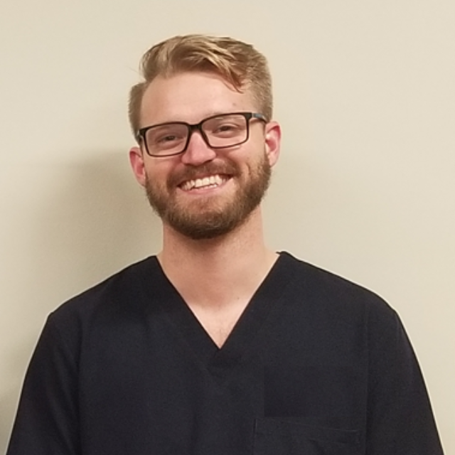
Our Echo and Stress Echo teams are made up of some of the region’s most highly qualified sonographers; individuals who have committed to specialized training in advanced diagnostic technologies including Microbubble UEA Echo. We are privileged to have these exceptional professionals with their highly sought-after skillset on our team. Their soothing bedside manner coupled with their excellent diagnostic acumen is among Cambridge Cardiac Care’s greatest assets.

Preparing for your appointment

Please follow these instructions the morning of your appointment:

  1. Enjoy a light meal 3-4 hours before your appointment.

  2. Stay away from caffeine. No coffee, tea, chocolate or soda pop. Please also avoid decaf products since they still have trace amounts of caffeine that can interfere with your test.

  3. Your healthcare provider may have directed you to stop certain medications the day before your appointment. Please DO NOT make any changes to your medication regimen without first speaking with your healthcare provider.

  4. Wear comfortable exercise clothes the day of your appointment as if you are going to the gym.

  5. Bring running shoes or sneakers with you to change into once are called into the testing room.

  6. Bring a list of all the medications you are currently taking.

  7. Expect for the appointment to last 60-90 minutes.

Medications may affect your test

Follow your healthcare practitioner’s instructions

Some patients may be required to discontinue taking certain medications. This does not, however, apply to all patients. So, please, do not stop any medications on your own without the express instructions of your healthcare provider. They should inform you at least 48 hours prior to your appointment if a temporary hold is required. On the day of your test, you may resume taking your medications immediately following the procedure.

Stress Echo

Patient Education

What you can expect, step by step.

  1. A technologist or sonographer will give you a consent form to sign and will explain the test to you. They will be happy to answer any questions you may have about the procedure. However, they will not be able to discuss the results of the test or what they see on the screen, in any way.

  2. You will be given privacy to change into a gown that will be provided to you. You will be asked to remove all upper body clothing and fasten the gown in the front. Making sure that you have running shoes or sneakers on and laced up, you will lie down on an examination table on your left side until the technologist returns.

  3. Adhesive electrodes will be applied to your chest to set up for ECG monitoring. The electrode sites will be cleaned with alcohol and possibly shaved to ensure an interference-free and continuous ECG recording.

  4. A technologist or nurse will insert an intravenous (IV) line into a vein in one of your arms.

  5. The sonographer will apply some gel to a small ultrasound probe (a transducer) and will place it on your chest, pressing down at times to take images of your heart at rest. Your resting blood pressure, heart rate, and ECG will also be recorded.

  6. Next, you will be asked to stand up and mount a treadmill that is located just steps away from the examination table.

  7. You will begin walking on the treadmill at a slow introductory pace that will be increased every 3 minutes until your target heart rate is reached.

  8. You will be monitored throughout the test. If a problem occurs, the technologist will stop the test right away. It is very important for you to tell the technologist if you experience any symptoms, such as chest pain, dizziness, unusual shortness of breath, or extreme fatigue.

  9. Once the treadmill portion of the test is complete, you will need to quickly lie back down on the examination table, on your left side for a second echocardiogram. This time, the technologist will capture images of your heart working hard after exercising on the treadmill.

  10. Your blood pressure, heart rate, and ECG will continue to be monitored for some time after the treadmill exercise. Your blood oxygen saturation may also be checked before and after the test and in some cases, a quick breathing test (spirometry test) may also be done before and after the treadmill portion of the stress echo.

  11. Data from the test will be reviewed by a cardiologist in a timely fashion. A report will then be sent to your healthcare provider.

  12. Following the test, you may resume your regular activities.

Why have I been referred for UEA Stress Echo?

Your healthcare provider may have many reasons to call for a UEA stress echocardiogram to be done at this time. Since it is a multifaceted test that gives comparative information of your heart at rest versus when it is taxed with exercise, it can be used to investigate the causes of exercise-induced symptoms like shortness of breath, lightheadedness or palpitations. It is also instrumental in the diagnosis of coronary artery disease (CAD) and chest pain (angina) symptoms. As a result, UEA stress echo can help predict the development of cardiovascular conditions including the elevated risk of a cardiac event, such as a heart attack, occuring in the future. The test also helps us determine the appropriate course of treatment in cardiac rehab & risk reversal by indicating the safe-zone of exercise based on fitness level and cardiac functioning.

What are the risks of “stressing” my heart during the stress test?

The UEA stress echo is meant to diagnose and prevent cardiac events from happening, not induce them. Complications during the stress portion of the test are extremely rare. Nevertheless, experienced medical staff who are trained to handle emergency situations are in attendance at all times. Patients are under constant cardiac monitoring during the test which means that it will be halted and appropriate medical attention will be received at the earliest sign.

How does echocardiography work?

Echocardiography is based on the same laws of nature that bats use to “see” in the dark (echolocation), that ships and fishermen use to navigate the deep sea (SONAR) and that obstetricians use to measure a baby in the womb (ultrasound). When a sound wave strikes an object, it bounces back, or echoes. By measuring these echo waves, it is possible to determine not only how far away an object is but also its size, shape and consistency.

In cardiology we use echocardiography ultrasound to detect changes in the appearance and functioning of the heart, its tissues, valves and vessels and to detect abnormal masses, such as tumors. This is facilitated by an ultrasensitive “wand” called a transducer which both sends sound waves into the body and receives the resulting echo. As the sound waves bounce off of internal organs, fluids and tissues, the transducer then receives the information and records tiny changes in the sound's pitch and direction. These signature waves are instantly measured and translated into an image that is displayed on the monitor.

Why do I need an IV for this test?

Our commitment to the safe and effective use of technology in the diagnostics and treatment of our patients is unwavering. To that end, we have invested in the advanced technology of microbubble UEA echocardiography. As a pain-free, biocompatible, non-invasive test that involves absolutely no radiation, it goes above and beyond safety standards while providing improved image resolution and diagnostic accuracy.

The IV is inserted at the beginning of the appointment, however, the microbubble UEA liquid is only administered through the IV at certain phases of the appointment when it is specifically needed to enhance the ultrasound image. When an ultrasound is conducted in the presence of this IV liquid, the sound waves emitted by the machine bounce off of the microbubbles giving high-resolution, real-time, moving images of the heart, its chambers, vessels, valves and their functions. Microbubble UEA echocardiography provides a huge advantage in the accuracy and reliability of test results.

Where do these microbubbles end up eventually? Since the bubbles consist of an inert, biocompatible gas, the body simply breathes them out through the lungs. It will be as undetectable to the patient as breathing out carbon dioxide and other naturally occurring gases with each out-breathe.

Is this test covered by OHIP?

Yes. OHIP covers your Stress Echo test provided it has been ordered for medical, diagnostic purposes. On some occasions, patients require Stress Echo monitoring for insurance or ministry of transportation assessment in which case, OHIP does not cover the test and the cost of the test is charged to the patient or their insurance directly.

Wait Times

Drag to see the table

Cardiac Test Routine Test Wait Time Urgent Test Wait Time
ABPM 2 weeks Same day
ECG 24 hours Same day
Echocardiogram 2 weeks 36 hours
Stress Test (Exercise / Treadmill) 2 weeks Same day
Stress Echocardiogram 2 weeks 24 hours
Holter Monitor - 24 Hour 2 weeks 24 hours
Holter Monitor - 48 Hour 2 weeks 24 hours
Holter Monitor – 72 Hour 2 weeks 48 hours
Holter Monitor - 2 Weeks 2 weeks 48 hours
Spirometry 24 hours Same day

The above wait-times indicate the standard benchmarks that Cambridge Cardiac Care strives to meet each and every day. Unforeseen circumstances and Public Health mandates, may effect these wait times. Rest assured that timely access is a driving force at CCC: we will get you in as soon as possible, based on your assessed level of urgency.